Korotkoff sounds in blood pressure measurements are the acoustic vibrations produced by turbulent blood flow as an inflated arm cuff gradually releases arterial compression. When a clinician wraps a cuff around your arm and listens through a stethoscope, they track the acoustic transitions that occur as the brachial artery shifts from complete occlusion to normal circulation. Russian physician Nikolai Korotkoff first identified these sounds in 1905, creating the foundation for modern manual auscultation. At BoringMagazine.Co, we explain how physical principles shape routine diagnostic tests. This guide provides general educational information about vascular acoustics rather than clinical advice. Anyone with questions about their personal readings should consult a qualified physician or healthcare provider.
How Arterial Occlusion Generates Blood Flow Acoustics
To understand why Korotkoff sounds occur, consider how blood moves through open vessels. Under resting conditions, blood travels through arteries in silent patterns known as laminar flow. Fluid travels in concentric layers, where the fastest stream flows down the center and fluid near the wall moves slower due to friction. Because these layers slide smoothly without colliding, laminar flow produces no sound on a stethoscope.
Sound emerges only when blood flow turns turbulent. Inflating a cuff above systolic pressure compresses the brachial artery against the humerus bone, creating arterial occlusion. As long as cuff pressure exceeds arterial pressure, blood cannot pass, and the stethoscope detects silence.
Acoustic vibrations begin when air vents from the cuff. As cuff pressure drops slightly below peak arterial pressure, blood spurts through the narrowed lumen during ventricular contraction. This high-velocity jet creates swirling vortices and pressure fluctuations that vibrate the arterial wall, transmitting rhythmic tapping sounds to the stethoscope.
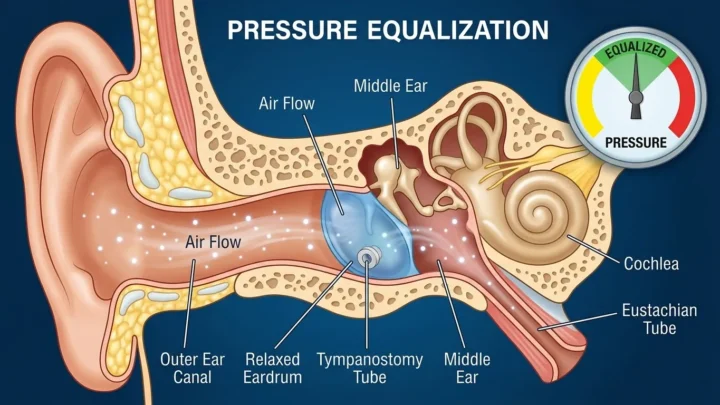
Fluid dynamics govern pressure variations throughout the human body. Just as mechanical fluid mechanics explain how ear tubes equalize middle ear pressure across the tympanic membrane, pressure differentials across compressed arterial walls drive vascular turbulence. You can explore related clinical concepts in our Health guides.
The Five Distinct Phases of Korotkoff Sounds
Clinical practice organizes the acoustic patterns heard during cuff deflation into five numbered phases, reflecting progressive changes in vessel diameter.
- Phase I: Clear, faint tapping sounds appear. These initial taps mark systolic blood pressure, occurring when peak cardiac force pushes the first spurts of blood through the compressed artery.
- Phase II: The sounds soften into a swishing murmur. Blood flows through the narrowed vessel for a longer fraction of each pulse cycle, generating acoustic turbulence.
- Phase III: The sounds become crisper, louder knocks. The artery opens wider during cardiac contraction, letting larger volumes of blood surge through at high speed.
- Phase IV: Sounds become distinctly muffled, soft, and blowing. This muffling occurs as external pressure approaches resting arterial pressure, keeping the vessel open across most of the cycle.
- Phase V: Complete silence returns. This disappearance marks diastolic blood pressure in adults. Once cuff pressure drops below resting arterial pressure, laminar flow resumes and vibrations stop.
What Systolic and Diastolic Readings Represent Mechanically
A blood pressure reading pairs two numbers, such as 120/80 millimeters of mercury (mmHg). These figures record two mechanical pressure balances identified by Phase I and Phase V sounds. Diagnostic organizations like the American Heart Association rely on these thresholds to define healthy cardiovascular parameters.
The upper value, systolic pressure, records peak force in the arterial tree when the left ventricle contracts. When a clinician hears Phase I, external cuff pressure matches this peak internal force. Even a tiny pressure drop allows a spurt of blood to break through, producing the first audible tap.
The lower value, diastolic pressure, measures baseline pressure while the heart rests between beats. As blood flows into peripheral capillary beds, arterial pressure declines to its minimum level. When cuff pressure drops below this resting minimum, the cuff no longer compresses the artery at any point during the heartbeat. The vessel stays round, laminar flow returns, and sound vanishes at Phase V.
In pregnancy or in young children, high blood flow velocity can create turbulence through open vessels, causing sound to persist down toward zero on the gauge. In those cases, clinicians use Phase IV muffling to identify diastolic pressure.
Step-by-Step Acoustic Detection with a Sphygmomanometer
Accurate auscultation requires a structured sequence with a manual sphygmomanometer and a stethoscope:
- Patient setup: The patient rests for five minutes with back support and feet flat. The arm rests on a table at heart level.
- Cuff fitting: The clinician selects a cuff whose bladder encircles 80 percent of the arm, wrapping it snugly two centimeters above the elbow crease.
- Pulse palpation: The clinician feels the radial pulse and inflates the cuff until the pulse stops, identifying the obliteration point to guide inflation.
- Stethoscope placement: The clinician places the stethoscope diaphragm lightly over the brachial artery below the cuff edge, avoiding excessive pressure.
- Controlled deflation: The clinician deflates the cuff at two to three millimeters of mercury per second, noting the reading at the first tap (Phase I) and at silence (Phase V).
Common Measurement Errors and the Auscultatory Gap
Several mechanical and technique mistakes can distort manual blood pressure readings.
Incorrect cuff sizing causes significant error. An undersized cuff requires excessive pressure to compress the artery, creating falsely high readings. An oversized cuff distributes force over too large an area, causing falsely low readings.
Rapid deflation also skews numbers. Releasing air faster than two to three millimeters of mercury per second causes the needle to drop past the true acoustic onset before the listener reacts, underestimating systolic pressure and overestimating diastolic pressure.
The auscultatory gap presents another clinical pitfall. This gap is a temporary silence between Phase I and Phase II, spanning 10 to 40 mmHg, before sounds return. It appears most often in older adults with arterial stiffness. If an examiner inflates the cuff into this silent zone without checking the pulse by touch first, they may mistake Phase II for Phase I, underestimating systolic pressure.
FAQ
What causes the tapping noise heard during a blood pressure test?
The tapping noise comes from turbulent spurts of blood striking the arterial wall as the compressed vessel opens briefly during cardiac systole. When cuff pressure drops slightly below peak arterial force, blood jets through the narrowed opening, generating vibrations that register as crisp clicks in a stethoscope.
Why do Korotkoff sounds stop at the diastolic pressure reading?
The sounds stop at the diastolic pressure point because blood pressure in the artery exceeds cuff pressure throughout the entire heartbeat, restoring smooth laminar flow. Once external pressure falls below resting arterial pressure, the vessel remains open, eliminating the turbulence required to produce acoustic vibrations.
Can digital blood pressure monitors hear Korotkoff sounds?
Most digital home blood pressure monitors do not hear Korotkoff sounds because they measure oscillometric pressure vibrations in the cuff rather than acoustic sound waves with a microphone. Electronic sensors record pressure oscillations transferred from the pulsing artery into the cuff bladder, using algorithms to calculate systolic and diastolic values.