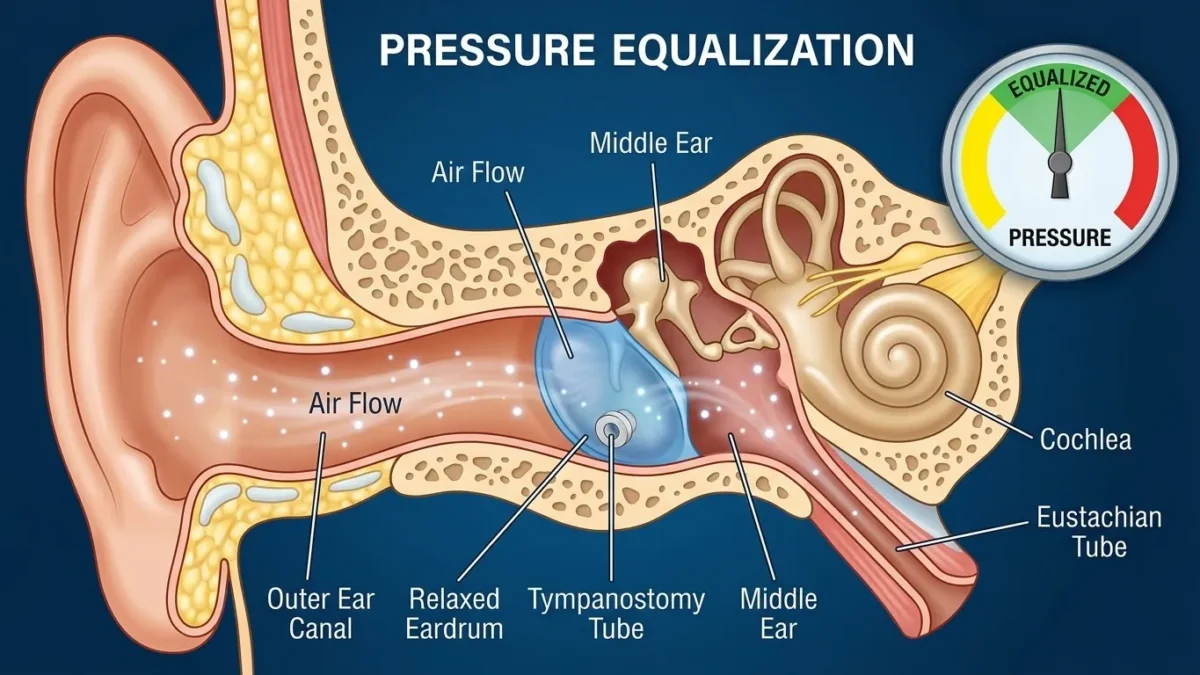
Ear tubes work in ears by providing a tiny ventilation channel across the eardrum that constantly equalizes air pressure between the middle ear and the outside world. When the body’s natural Eustachian tubes fail to open, this small cylinder prevents a vacuum from forming and allows trapped fluid to escape. At BoringMagazine.Co, we examine everyday medical devices to show how simple physical mechanisms solve complex anatomical problems.
The Mechanics of Equalizing Pressure Across the Eardrum
The human ear depends on balanced air pressure to transmit sound clearly. Sound waves travel down the ear canal and strike the tympanic membrane, commonly called the eardrum. The eardrum vibrates and transfers energy through three tiny bones in the middle ear: the malleus, incus, and stapes. For the membrane to vibrate freely, air pressure inside the middle ear cavity must match atmospheric pressure outside the head.
Tissues in the middle ear constantly absorb trapped air. If fresh air does not replace these gases, internal pressure drops. This drop turns the middle ear into a low-pressure sealed space. The higher ambient pressure outside pushes the flexible eardrum inward, stretching it tight and restricting the movement of the tiny bone chain.
A tympanostomy tube creates an artificial airway directly through the tympanic membrane. The tube is a hollow spool made from silicone or fluoroplastic. Because the core remains open, ambient air flows freely between the ear canal and the middle ear space. This physical channel balances pressure on both sides of the eardrum, allowing the membrane to regain normal flexibility and vibrate without resistance.
Why Natural Ventilation Fails: Eustachian Tube Dysfunction
Under normal conditions, the body relies on the Eustachian tube rather than the eardrum for ventilation. The Eustachian tube is a narrow canal running from the middle ear to the back of the throat. Muscles open this canal whenever a person swallows or yawns, which introduces fresh air and drains secretions.
When the Eustachian tube swells shut or fails to open, doctors call the problem Eustachian tube dysfunction. Several factors block this passage:
- Immature anatomy, because young children have shorter, flatter tubes that drain poorly.
- Enlarged adenoid tissue that blocks the entrance of the tube in the nasopharynx.
- Mucosal swelling caused by viral infections or an IgE-mediated allergy.
- Muscular weakness that prevents the tube from opening during swallowing.
When a blockage persists, negative pressure pulls fluid out of surrounding tissues into the empty middle ear cavity. To explore more biological explanations behind immune responses and inflammation, read our Health guides. Without an open airway, the fluid turns thick, leading to chronic middle ear effusion.
The Placement Procedure and Key Tube Designs
Placing an ear tube involves a brief outpatient procedure called a myringotomy with tube insertion. Ear specialists perform the surgery under a microscope. Young children receive brief general anesthesia, while older patients can undergo the procedure with local numbing drops.
The surgeon completes four direct steps:
- Examines the eardrum and clears excess wax from the canal.
- Makes a tiny incision in the lower section of the eardrum.
- Suctions out trapped fluid or mucus from the middle ear space.
- Inserts the small tube into the incision, seating its rims like a grommet.
Surgeons select tube types based on the expected duration of treatment:
- Short-term grommets: These spool-shaped tubes stay in place for six to eighteen months. The healing eardrum sheds skin cells outward, pushing the tube into the canal naturally.
- Long-term tubes: Designs such as Goode T-tubes feature wide, flexible flanges that resist extrusion. They stay in place for two or more years in patients with persistent structural collapse.
Clinical Examples and Patient Scenarios
Tympanostomy tubes solve distinct physical problems across different age groups. Real clinical situations show how equalizing pressure restores hearing and prevents infection cycles.
Consider a toddler with recurrent acute otitis media who experiences four infections in five months. Each episode brings intense ear pain, fever, and oral antibiotics. The child’s eardrum bulges outward under fluid pressure. Placing a tube vents the cavity and keeps it dry. If bacteria enter later, antibiotic drops reach the space directly while fluid drains freely without painful pressure buildup.
Another case involves a preschool child with silent fluid behind both eardrums lasting four months. Without fever or pain, the thick fluid dampens vibrations and causes mild conductive hearing loss. The National Institute on Deafness and Other Communication Disorders notes that middle ear fluid muffles sound and can affect early speech development. Inserting tubes drains the fluid and restores normal sound conduction immediately.
Adults also benefit during frequent air travel or hyperbaric oxygen therapy. Inability to equalize middle ear pressure during rapid altitude changes causes severe barotrauma. A tube equalizes pressure automatically and protects the eardrum from tearing.
Common Misconceptions About Tympanostomy Tubes
Because ear tubes sit out of sight, several misconceptions persist regarding their function and safety.
One misconception is that ear tubes leave a permanent hole in the eardrum. In most cases, the tympanic membrane closes on its own as it pushes the tube outward. New skin cells knit the slit closed behind the tube. Permanent perforations occur in only a tiny fraction of patients and can be repaired if necessary.
Another belief is that children with tubes must avoid water entirely. Modern clinical guidelines show that clean bathwater and surface swimming in treated pools rarely enter standard tubes due to liquid surface tension. Ear protection is primarily required for deep diving or swimming in untreated lake water.
Some people also believe tubes stop infections from happening. Tubes do not alter the immune system or kill bacteria. Instead, they prevent fluid from remaining trapped under pressure. When an infection occurs, it produces drainage rather than painful pressure.
This article provides educational information rather than specific clinical advice. Anyone experiencing ear pain, hearing changes, or repeated infections should consult a qualified physician for an evaluation.
FAQ
Here are clear answers to frequent questions about tympanostomy tubes and middle ear pressure.
How long do ear tubes stay inside the ears?
Ear tubes usually stay inside the eardrum for six to eighteen months before falling out naturally. As the eardrum heals, migrating skin cells loosen the tube until it drops into the ear canal. The tube often falls out unnoticed or is discovered during a routine checkup.
Can patients feel ear tubes after placement?
Patients cannot feel ear tubes after placement because the tiny cylinders sit in the eardrum without contacting sensitive ear canal walls. The light synthetic material creates no sensation of weight or pressure once the initial incision heals. Children return to normal activities right after recovery.
Does drainage through the ear tube mean the tube is failing?
Drainage through the tube means the device is working properly by allowing fluid to escape instead of building pressure behind the eardrum. When a cold causes middle ear secretions, fluid flows outward through the tube into the canal. Physicians treat this drainage with targeted antibiotic ear drops.