Messenger RNA instructions work in cells by carrying temporary genetic blueprints from DNA to ribosomes, which read the sequence and assemble specific amino acids into proteins. Every human cell relies on this transcription and translation pathway to build enzymes, structural fibers, and signaling molecules necessary for survival. At BoringMagazine.Co, we focus on explaining everyday biological processes through clear, observable mechanics. Understanding how cells read and dismantle these molecular messages clarifies how tissues repair themselves and how modern medical interventions train the immune system.
How Cells Read mRNA Instructions
Messenger RNA, or mRNA, is a single-stranded molecule composed of four nucleotide bases: adenine, uracil, cytosine, and guanine. In human cells, genomic DNA stays protected inside the nucleus. When a cell needs a specific protein, it transcribes the gene into an mRNA strand. This transcript receives a protective five-prime cap and a polyadenylated tail before exiting into the cytoplasm.
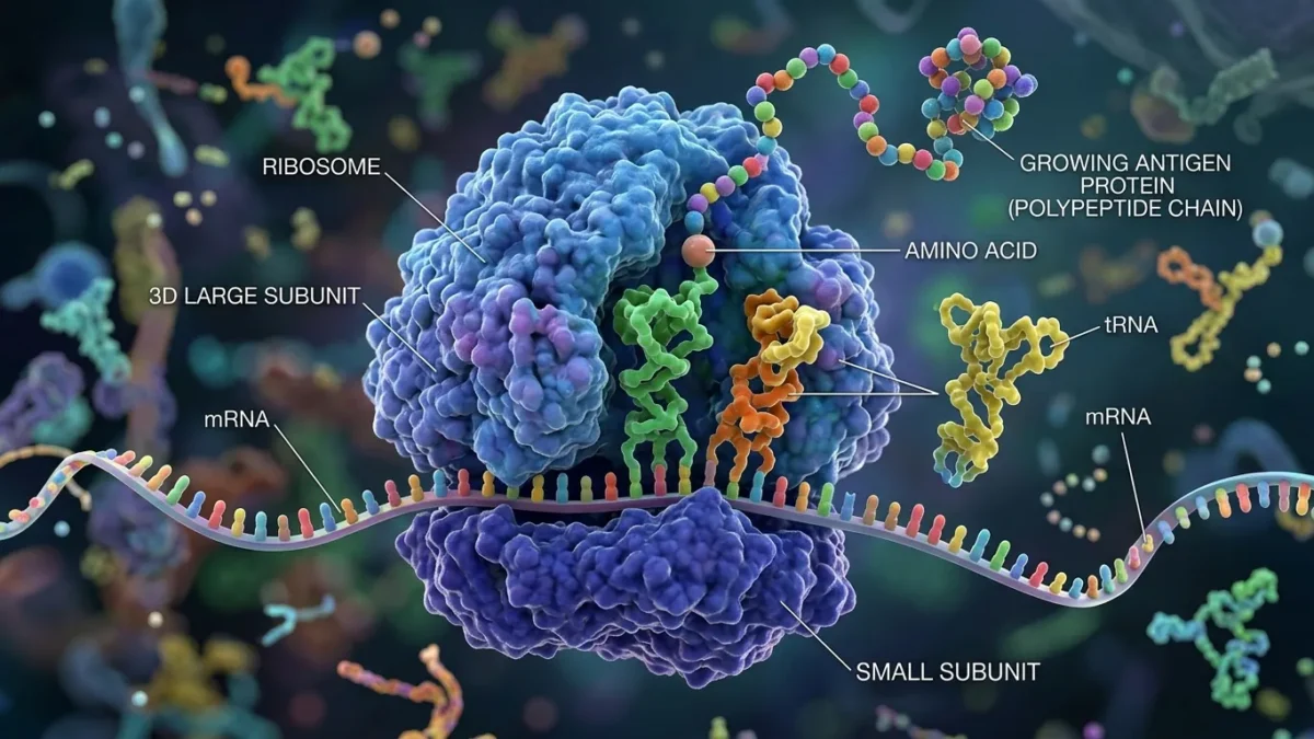
In the cytoplasm, mRNA meets ribosomes. Ribosomes consist of ribosomal RNA and proteins structured into small and large subunits. The information on the mRNA is organized into three-nucleotide triplets called codons, each coding for a specific amino acid or a stop command. Transfer RNA molecules act as physical adaptors. Each tRNA features an anticodon matching an mRNA codon and carries the corresponding amino acid. According to the National Institutes of Health, this codon-anticodon pairing ensures that ribosomes assemble amino acids in the exact sequence specified by the genetic code.
The Three Main Steps of Ribosomal Translation
Ribosomal translation transforms the linear sequence of nucleotides into a functional protein through three stages: initiation, elongation, and termination.
- Initiation: The small ribosomal subunit attaches to the five-prime cap of the mRNA strand and scans for the start codon, usually adenine-uracil-guanine. An initiator tRNA carrying methionine binds to this site. The large ribosomal subunit then docks on top, assembling a functional ribosome with active aminoacyl, peptidyl, and exit binding sites.
- Elongation: The ribosome moves along the mRNA molecule one codon at a time. An incoming tRNA enters the aminoacyl site, pairing its anticodon with the mRNA codon. The ribosome forms a peptide bond between the growing peptide chain and the new amino acid. As the ribosome steps forward, the empty tRNA exits, making room for the next match. This cycle repeats until the full protein chain forms.
- Termination: Protein synthesis concludes when the ribosome reaches a stop codon: uracil-adenine-adenine, uracil-adenine-guanine, or uracil-guanine-adenine. Release factor proteins bind to these codons rather than a tRNA. The ribosome releases the completed polypeptide chain and detaches from the mRNA. The subunits separate, ready to translate another sequence.
Natural RNA Degradation and Cellular Cleanup
A fundamental feature of cellular biology is that mRNA messages are temporary. Without natural degradation, cells would produce proteins unchecked and disrupt normal metabolism. Cells rely on strict cleanup mechanisms to dismantle mRNA after translation.
An mRNA molecule lifespan depends heavily on its polyadenylated tail, a stretch of adenine nucleotides at the three-prime end. Cytoplasmic deadenylase enzymes steadily shorten this tail. Once the tail degrades past a critical threshold, decapping enzymes remove the protective five-prime cap, exposing the RNA strand to cellular ribonucleases.
Ribonucleases, or RNases, are cytoplasmic enzymes that cleave the chemical bonds holding the RNA chain together. They break the strand into individual ribonucleotides. The cell recycles these subunits to build new RNA molecules or convert them into cellular energy. Thanks to this routine enzymatic clearance, mRNA has a functional half-life of only a few hours, guaranteeing that protein synthesis ceases promptly.
Synthesizing Antigen Proteins for Immune Defense
The same ribosomal machinery that produces structural proteins can also synthesize antigen proteins. In immunology, an antigen is a molecular structure that triggers an adaptive immune response. When a viral infection occurs, or when an engineered mRNA sequence enters a cell inside a protective lipid envelope, ribosomes do not distinguish between native and introduced instructions. They translate the template using the cellular pool of amino acids.
In modern mRNA vaccines, the sequence codes for a harmless viral surface feature, like a spike glycoprotein. Ribosomes translate the spike protein in the cytoplasm. Cellular transport systems then move the finished antigen toward the cell surface, where major histocompatibility complex molecules display fragments to the outside environment.
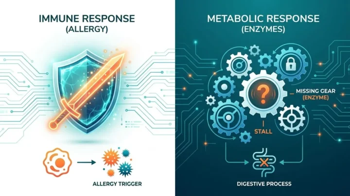
Circulating immune sentinels, such as dendritic cells and lymphocytes, identify these displayed fragments as foreign. This recognition prompts B lymphocytes to produce specific neutralizing antibodies and primes T lymphocytes to destroy infected cells in the future. Just as we detail in our Health guides, learning how the body differentiates normal cellular components from outside triggers is key to understanding immune protection. For instance, exploring the physiological difference between an allergy and an intolerance shows how sensitive immune receptors are to specific protein structures. Readers with specific questions regarding immunization schedules or immune deficiencies should consult a qualified physician or healthcare provider for individualized medical advice.
Common Misconceptions About mRNA Function
Public discussions regarding mRNA technology frequently involve misunderstandings about cell anatomy and molecular genetics. Reviewing basic biology helps clarify what mRNA can and cannot do inside the human body.
- mRNA does not alter genomic DNA: Human DNA remains protected inside the nucleus by a double membrane. Messenger RNA operates solely in the cytoplasm. Because human cells lack reverse transcriptase enzymes, an mRNA strand cannot copy itself into DNA or insert into chromosomes.
- mRNA does not accumulate permanently: Native and foreign mRNA strands break down quickly once translation finishes. Cytoplasmic RNases dismantle the instructions within hours or days, leaving only the synthesized protein and the resulting immune memory.
- mRNA does not generate live pathogens: Therapeutic mRNA carries code for a single isolated antigen protein rather than a full virus. Without the complete viral genome, a cell cannot assemble an infectious particle.
- mRNA does not assemble antibodies directly: Messenger RNA instructs ribosomes to build target antigens, not antibodies. The host immune system subsequently detects these antigens and manufactures its own antibodies.
FAQ
Where does mRNA translation occur inside the cell?
Translation occurs in the cytoplasm of the cell, where ribosomes bind to mRNA strands away from the cell nucleus. After leaving the nucleus, the mature mRNA moves into the fluid cytoplasm or attaches to the endoplasmic reticulum for translation.
How long does mRNA remain active before breaking down?
Cellular mRNA molecules typically persist for only a few hours to several days before cellular enzymes degrade them completely. Structural elements like the polyadenylated tail and local ribonuclease concentrations determine the exact speed of breakdown.
Can mRNA enter the cell nucleus to alter human DNA?
Synthetic or cellular mRNA cannot enter the cell nucleus or alter DNA because it lacks the chemical signals required to cross the nuclear membrane and cannot integrate into genomic material. Human cells also lack the reverse transcriptase enzymes required to turn RNA into DNA.